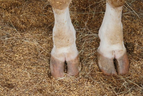
PATHOLOGY DESCRIPTION
Lameness is the third most important problem on many modern dairy farms after mastitis and reproductive failure. The considerable economic losses are attributable to the cost of treatment, decreased milk production, decreased reproductive performance and increased culling. The incidence of lameness has steadily increased over the past 20 years and on some farms over half of the animals become lame at least once each year. Lameness is a symptom related to several diseases; with multifactoriel origins. nutritional mismanagement, wet environment, abrasive or slippery floor surfaces, stall comfort and design and health events causing production of poor quality horn. We will focus on the 3 main infectious diseases responsible of lameness usually resulting from poor hygiene.
FOOTROT
• Description
Footrot has a worldwide distribution and is usually sporadic but may be endemic in intensive beef or dairy cattle production units. The incidence varies according to weather, season of year, grazing periods and housing system. On average, footrot accounts for ~15% of claw diseases.
• Origin
Injury to the interdigital skin provides a portal of entry for infection. Maceration of the skin by water, feces and urine may predispose to injuries. Fusobacterium necrophorum is considered to be the major cause of footrot. It can be isolated from feces, which may explain why control is difficult. Other organisms, such as Staphylococcus aureus, Escherichia coli, Arcanobacterium (Actinomyces) pyogenes and possibly Bacteroides melaninogenicus, can also be involved.
• Symptoms
Footrot is a subacute or acute necrotic infection originating from a lesion in the interdigital skin. Pain, severe lameness, fever, anorexia, loss of condition and reduced milk production are major signs of the disease.
INTERDIGITAL DERMATITIS
• Description
Interdigital dermatitis is a low-grade infection of the interdigital epidermis that causes a slow erosion of the skin with discomfort but no lameness unless the lesion becomes complicated. It is seen worldwide but is most prevalent under poor hygienic conditions in intensive dairy production. Morbidity is usually high in housed animals, particularly toward the end of the winter. When animals in such herds are examined, it is not unusual for 100% to have lesions of varying degrees of severity. The prevalence of heel horn erosion may increase in herds that have a high prevalence of interdigital dermatitis, suggesting a close relationship between the 2 diseases.
• Origin
Interdigital dermatitis is caused by a mixed bacterial infection, but Dichelobacter nodosus has been considered to be the most active component. The disease is most commonly seen when humidity is high, in temperate climates and under poor hygienic conditions, especially in housed dairy cattle. The source of the infection is the cow itself and the infection spreads from infected to non-infected animals through the environment. Dnodosus cannot survive >4 days on the ground but can persist in filth that is caked onto the claws.
• Symptoms
The bacteria invade the epidermis, but the organisms do not penetrate to the dermal layers. As the condition progresses, the border between the skin and soft heel horn disintegrates, producing lesions similar to ulcers or erosions. At this stage, the lesions cause discomfort.
DIGITAL DERMATITIS
• Description
Digital Dermatitis, also called Mortellaro disease, is a highly contagious, erosive and proliferative infection of the epidermis proximal to the skin-horn junction in the flexor region of the interdigital space. Morbidity within a herd can be >90%. It can affect any breed or age group, although young animals with a poor immune response are most susceptible. It spreads rapidly from newly acquired animals, or it may be introduced by any mechanical vector, eg, boots or hoof trimming instruments. The prevalence is highest in the fall and winter and is lowest if the animals are pastured.
• Origin
Deep in the epidermis of erosive/reactive lesions, 2 types of spirochetes can be demonstrated. One is a long, spiral, filamentous organism 12 × 0.3 μm and the other is a shorter, thicker spirochete 5-6 × 0.1 μm. However, it is thought that there is a multifactorial etiology with multiple organisms involved. Dichelobacter nodosus is likely to be implicated. Strong circumstantial evidence suggests that a virus plays a part in the pathogenesis of the disease, but to date, none has been isolated.
• Symptoms
Two main types of lesions are seen, one is erosive/reactive, the other is proliferative or wart-like. Both forms cause varying degrees of discomfort and may give rise to severe lameness. Sometimes, one particular form predominates in one area, but both forms can be seen in the same animal. The 2 forms likely represent different stages of the disease process. Some of the variation may be due to concurrent interdigital dermatitis.
COST
• Decrease milk production.
• Reproduction consequence.
• Extra replacement costs.
• Treatment costs.
